Branch Retinal Vein Occlusion (BRVO)
Retinal vein occlusions occur when there is a blockage of veins carrying blood with oxygen and nutrients to the nerve cells in the retina. A blockage in the retina’s main vein is referred to as a central retinal vein occlusion (CRVO), while a blockage in a smaller vein is called a branch retinal vein occlusion (BRVO).
Symptoms- Blurred vision
- Floaters
- If the affected area is not in the centre of the eye, BRVO can go unnoticed with no symptoms.
Most BRVOs occur at an arteriovenous crossing—an intersection between a retinal artery and vein. These vessels share a common sheath (connective tissue), so when the artery loses flexibility, as with atherosclerosis (hardening of the arteries), the vein is compressed.
The narrowed vein experiences turbulent blood flow that promotes clotting, leading to a blockage or occlusion. This obstruction blocks blood drainage and may lead to fluid leakage in the center of vision (macular edema) and ischemia—poor perfusion (flow) in the blood vessels supplying the macula.
Risk factorsThe common risk factors for BRVO are:
- Uncontrolled high blood pressure
- Being overweight or obese (increased body mass index)
- Cardiovascular (heart) disease
- Diabetes
- Glaucoma
- In younger patients who suffer BRVO, an abnormal tendency to develop blood clotting is also possible.
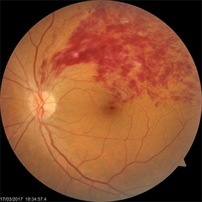
Most often, BRVO is diagnosed by an eye exam that shows retinal hemorrhage (blood vessels leaking into the retina), thickened and twisted blood vessels, and retinal edema (swelling with fluid).
Two types of retinal imaging tests aid the diagnosis of BRVO:
- Fluorescein angiography (FA)
- Optical coherence tomography (OCT)
FA provides images of fluid leaking from damaged or abnormal retinal vessels, demonstrating:
- Venous stasis (congestion and slowing of circulation)
- Edema (swelling with fluid)
- Ischemia (inadequate blood supply) or
- Retinal neovascularization (abnormal growth of new blood vessels in the retina)
OCT provides detailed images of the central retina, allowing detection of macular edema and fluid outside the macula.
TreatmentTreatment begins with identifying underlying risk factors and treating them. Risk factors are assessed using several methods:
- Blood pressure monitoring
- Determining if blood cholesterol or lipid levels are elevated
- Blood tests, if appropriate, to determine if there is an abnormal tendency to form blood clots
Eye treatment is aimed at treating retinal complications rather than at trying to relieve the blockage itself.
Macular edema, the main reason for visual loss from BRVO, is often treated with intraocular (in-the-eye) injections of anti-VEGF drugs designed to stop the growth of abnormal new blood vessels in the eye and decrease leakage. Local anesthetic eye drops are given before the injections to numb the eye and minimize discomfort.
There are currently 3 anti-VEGF drugs:
- Avastin® (bevacizumab®)
- Lucentis® (ranibizumab®)
- Eylea® (aflibercept®)
In several large clinical studies, all 3 of these anti-VEGF drugs have demonstrated good results, with over 50% of patients enjoying significant visual improvement. The use of these drugs may require frequent retreatment, but injection schedules are determined on a case-by-case basis.
Laser treatment may be used along with anti-VEGF therapy in hard-to-treat cases. Laser therapy for macular edema involves applying light laser pulses to the macula in a grid pattern. In a large multi-center clinical trial, after 3 years of follow up, this treatment showed improvement of vision in approximately two-thirds of patients.
Intraocular injections of steroids are another potential treatment for eyes that don’t respond to anti-VEGF drugs. A clinical trial that evaluated steroid treatment using a slow-releasing steroid implanted in the eye (dexamethasone or Ozurdex®), showed that approximately 30% of BRVO patients enjoyed significant visual improvement following treatment.